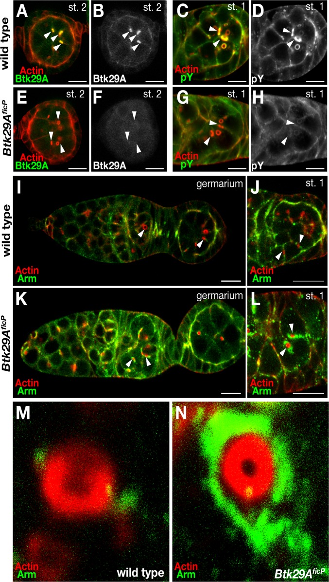
Fig 3. Effect of the Btk29A ficP mutation on the localization of Btk29A, Arm and phosphotyrosine.
The panels show the stage 2 (A, B, E and F) and the stage 1 (C, D, G and H) egg chambers from wild-type (A—D) and Btk29A ficP mutant (E—H) ovaries doubly stained with phalloidin for actin (red in A, C, E and G) and the anti-Btk29A antibody (green in A and E) or the anti-phosphotyrosine antibody (green in C and G). The images of antibody staining are also shown in black and white in (B), (D), (F) and (H) to aid in comparisons of the localization and abundance of Btk29A or phosphotyrosine between the wild-type and Btk29A ficP mutant egg chambers. Btk29A is highly enriched around ring canals and cell-cell contact regions in the wild-type chambers, whereas it is barely detectable in the Btk29A ficP mutant chambers. (I—N) Arm (green) is localized along the cell-cell contact regions and ring canals in the wild-type (I) and Btk29A ficP mutant (K) germaria. Actin is visualized by phalloidin staining (red). The entire stage 1 (J and L) and close-up views of ring canals in stage 1 (M and N) of the wild-type (J and M) and Btk29A ficP mutant (L and N) germaria are shown. Some of the ring canals are marked with arrowheads in (A—L). Note the marked accumulations of Arm in the regions surrounding the ring canals in Btk29A ficP mutants. Scale bars: 10 μm.

