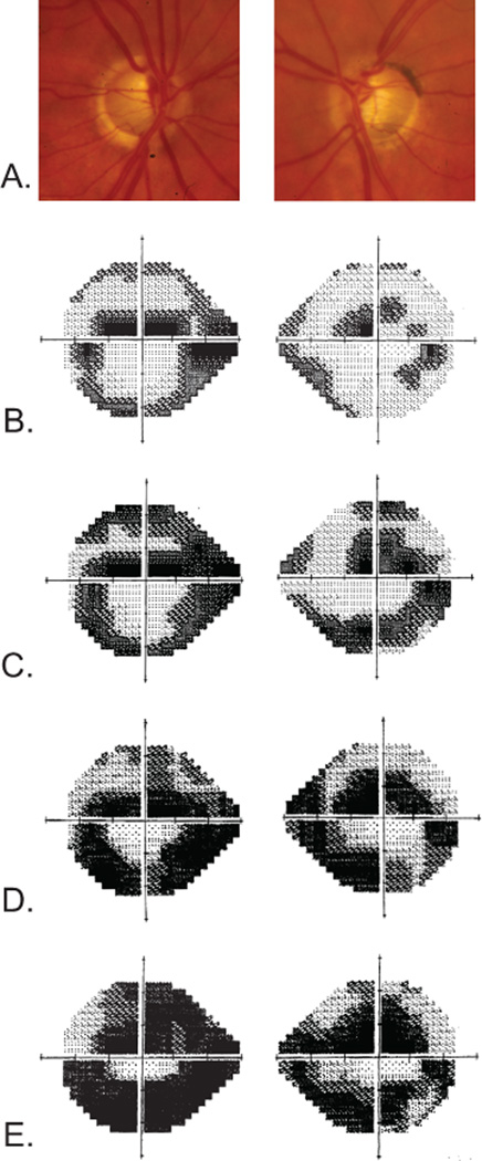
Figure 2. Patient GGR-590-1 clinical data.
A, Disc photographs at 56 years of age that demonstrate significant cupping. Humphrey visual field tests (24-2 Swedish Interactive Thresholding Algorithm Standard) performed at 53 years of age (B), 56 years of age (C), 59 years of age (D), and 64 years of age (E) demonstrate progressive glaucomatous visual field loss despite maximum intraocular pressure of 12 mm Hg in both eyes.

