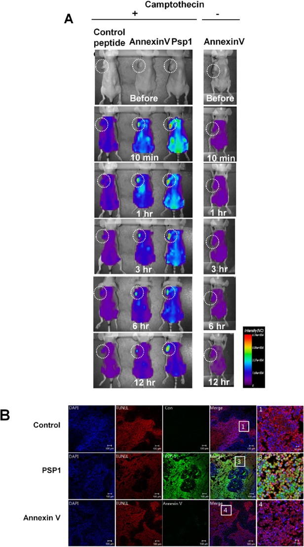
Fig 4. In vivo imaging of tumors after apoptosis induction.
(A) Cy7.5-labelled PSP1peptide, control peptide, or annexin V was systematically injected into the tail vein of tumor-bearing nude mice treated with camptothecin 24 hrs before injection. The same amount of annexin V was also injected into non-camptothecin-treated tumor-bearing mice as a control. The homing of the peptides was examined after the indicated times using an optical imaging system. The white dotted circles indicate the tumors. (B) Histological examination of tumor tissues after homing of PSP1 and annexin V, performed under a fluorescence microscope. Tumor cell apoptosis was observed by TUNEL staining.

