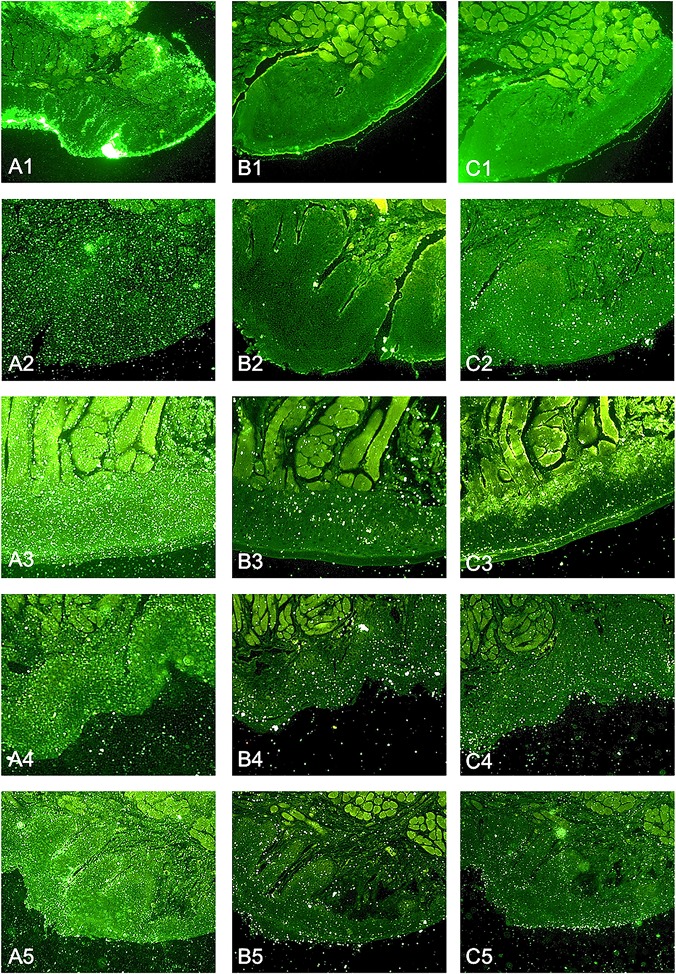
Fig 5. Representative fluorescence microscopy images of FITC labeled P. gingivalis adherent to murine oral mucosa sections from four independent experiments; data sets 4 and 5 originate as technical replicates from the same experiment to indicate intraassay reproducibility A 1–5: untreated control B 1–5: positive control, pretreated with 5 mM TLCK for 90 minutes C 1–5: RA1 100 μg/mL (preincubation of bacteria for 90 minutes).
Magnification: 100 ×. Images are equalized in brightness, contrast and fluorescence intensity.

