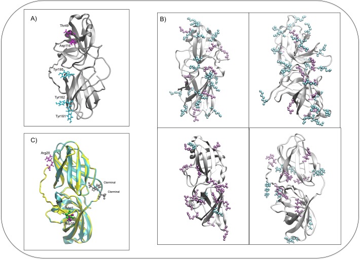
Fig 3. Structure of ScExlx1 and comparison with previously crystallized expansins.
A) Three-dimensional model proposed for ScExlx1. Most conserved amino acids between plant and microbial expansins in D1 are depicted in magenta (Thr-49 and Asp-116). Sugar-binding residues in D2 are showed in cyan (Tyr-160, Tyr-161 and Tyr-195). B) Three-dimensional models showing the positive charged amino acids (Arg+Lys) between different EXLX proteins reported previously and ScExlx1 (Lysine is depicted in cyan and Arginine is depicted in magenta). Top-left, PDB: 3D30. Top-right, PDB: 2HCZ. Bottom-left, PDB: 4JCW. Bottom-right, ScExlx1. C) BsExlx1 (cyan model) and ScExlx1 (yellow model) superimposed, showing an N-terminal extension in ScExlx1 that it is absent in BsExlx1. Amino acids depicted in silver, C-terminal in both proteins. Amino acid in green, Met-1 of BsExlx1. Amino acids in purple, Arg-20 and Asp-38 depicting the N-terminal extension in ScExlx1).

