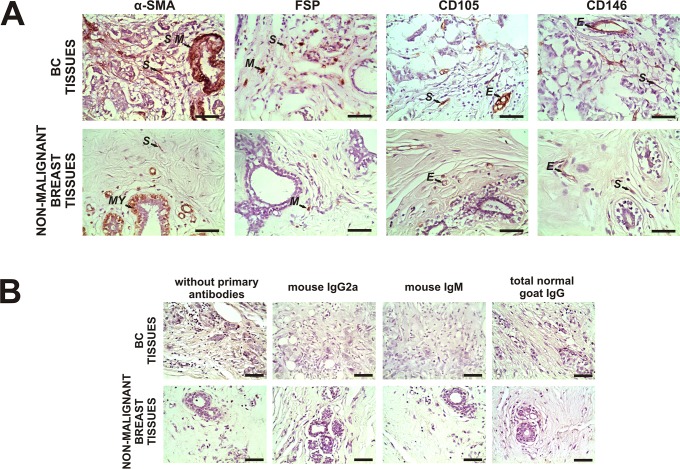
Fig 1. Expression of α-SMA, FSP, CD105 and CD146 in malignant and non-malignant breast tissues.
A Representative immunohistochemistry staining for α-SMA, FSP, CD105 and CD146 in stromal cells of primary tumor tissues from breast cancer (BC) patients and non-malignant breast tissues. Reactions were evaluated in spindle-shaped stromal cells, not associated with the vasculature. The arrows show positive staining of evaluated stromal cells (S) and smooth muscle cells (S M), myoepithelial cells (MY), tissue-associated macrophages (M) and endothelial cells (E) used as internal positive controls as previously described. B No staining was observed in both types of tissues when we incubated them without primary antibodies and with irrelevant mouse IgG2a (for α-SMA), irrelevant mouse IgM (for FSP) or total normal goat IgG (for CD105 and CD146) as negative controls. Nuclei were counterstained with hematoxylin (purple). Original magnification A and B: 400×. The scale bars represent 50 μm.

