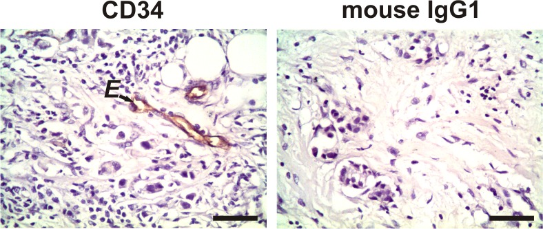
Fig 2. Expression of CD34 in tumor stromal cells.
Representative negative staining for CD34 in the evaluated spindle-shaped stromal cells, not associated with the vasculature, of primary tumor tissue from a breast cancer patient. The arrow shows an example of CD34-positive staining only in the endothelial cells (E). No staining was observed in the tissues when we incubated them with irrelevant mouse IgG1 as a negative isotype control. Nuclei were counterstained with hematoxylin (purple). Original magnification: 400×. The scale bar represents 50 μm.

