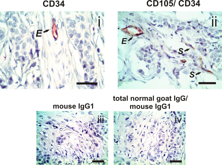
Fig 3. Expression of CD105 and CD34 in tumor stromal cells.
i Single immunohistochemistry for CD34 (detected by red chromogen) shows a representative example of CD34-positive staining in the endothelial cells (E). ii Double immunohistochemistry for CD105 and CD34 (detected by brown and red chromogen, respectively) shows a representative example of co-staining of CD105 and CD34 in the endothelial cells (E) and CD105-positive staining only in evaluated stromal cells (S) of primary tumor tissue from a breast cancer patient. No staining was observed in the tissues when we incubated them with irrelevant mouse IgG1 (for CD34) or sequentially incubated with total normal goat IgG and irrelevant mouse IgG1 (for CD105/ CD34) as negative isotype controls (iii and iv; respectively). Nuclei were counterstained with hematoxylin (purple). Original magnification: 400×. The scale bars represent 50 μm.

