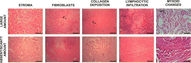
Fig 4. Histological features of primary breast tumor stroma as determined by hematoxylin and eosin staining.
This picture is an example of samples with a large amount and absent/scanty amount of stroma, fibroblasts, collagen deposition, lymphocytic infiltration and myxoid changes. The arrows show tumor fibroblasts (F), collagen deposition (C), lymphocytic infiltration (L) and myxoid changes (M C). Original magnification: 100×. The scale bars represent 200 μm.

