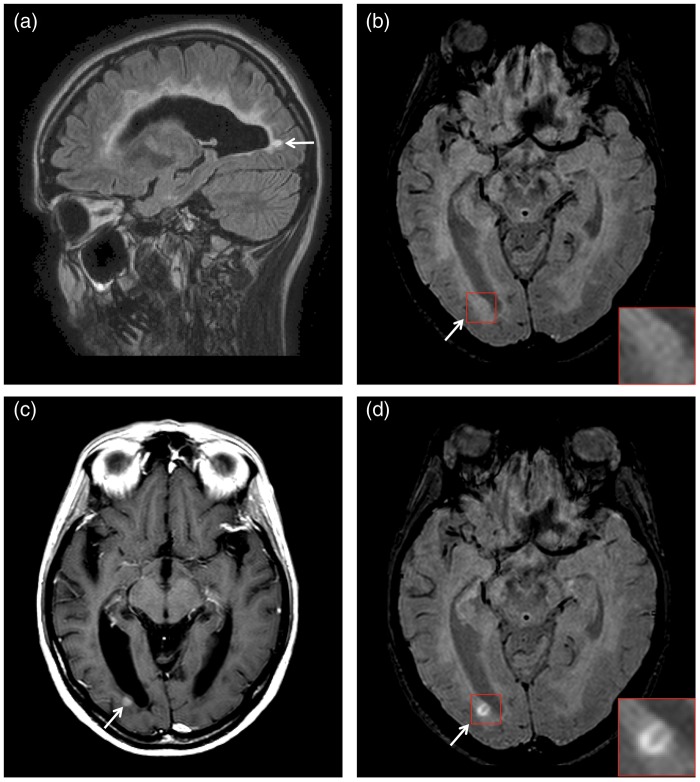
Fig. 2.
Occipital periventricular WM lesion (arrows) appearing hyperintense on (a) the sagittal pre-CM FLAIR and (b) the pre-CM axial SWI (magnified view). (c) The lesion appears visibly contrast-enhancing on the post-CM axial T1W SE and (d) the post-CM axial SWI. The post-CM SWI shows a linear shaped area of signal hypointensity in the lesion center, a parenchymal vein, which is not visible on the pre-CM SWI (magnified view).

