FIG 2.
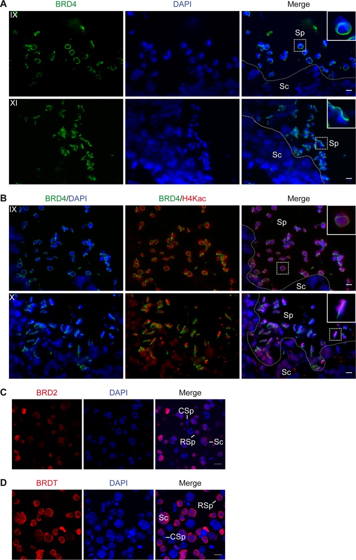
BRD4 forms a ring around the spermatid nucleus as histones become hyperacetylated. (A) Indirect IF assay of cryosectioned mouse testis tissue shows that BRD4 (green) forms a ring around the early (top) to late (bottom) elongating spermatid nucleus (DAPI-stained DNA in blue). The ring is absent from all nonspermatid cell types, such as spermatocytes (Sc). (B) Indirect IF assay of cryosectioned mouse testis tissue shows that the BRD4 ring (green) forms at the onset of histone H4 hyperacetylation (red) in the nuclei (DAPI-stained DNA in blue) of early (top) to late (bottom) elongating spermatids. The stage of spermatogenesis is shown in the upper left corner of each panel. Separation of spermatocytes (Sc) and spermatids (Sp) within the seminiferous tubule is indicated with a dotted line. The inset shows a 3× magnification of the spermatids outlined with a dotted square. (C and D) Indirect IF assay of a mixed population of spermatogenic cells shows that BRD2 and BRDT (red) are diffusely localized in the nuclei (DAPI-stained DNA in blue) of spermatocytes (Sc) and round spermatids (RSp) but not condensing spermatids (CSp). Scale bars, 10 μm.

