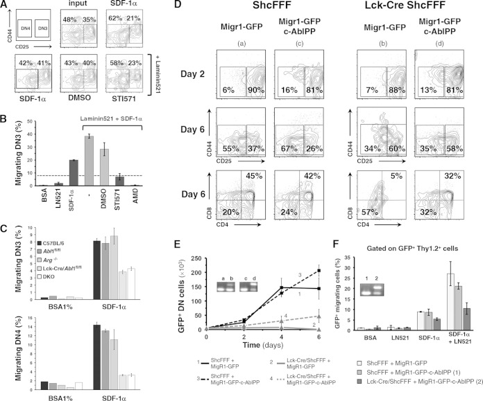
FIG 8.
Expression of constitutively active c-Abl partially rescues thymocyte differentiation, proliferation, and migration. (A and B) Purified DN thymocytes from C57BL/6 mice were pretreated with STI571 or AMD3100 and assayed for migration to SDF-1α in a transwell assay. Bar graphs show the percentage of migrated DN3 cells as a fraction of total cell input ± SD. DMSO, dimethyl sulfoxide; AMD, AMD3100. (C) Thymocytes from control abl1fl/fl, arg−/−, Lck-Cre/abl1fl/fl, and double-knockout (DKO) mice (three mice for each group) were assayed for SDF-1α-dependent chemotaxis, and the fraction of migrated cells is depicted as a percentage of DN3 or DN4 thymocyte input. (D) Differentiation of sorted DN3E thymocytes from ShcFFF and Lck-Cre/ShcFFF mice that were retrovirally transduced with c-AblPP or with empty GFP-expressing control plasmid and seeded on OP9-DL1 stromal cells (the analysis was performed on day 5). Dot plots were first gated on Thy1.2+ GFP+ cells. (E) Absolute numbers of total DN thymocytes in the coculture on days 2, 4, and 6. Insets show stop sequence deletion (detected via PCR) from genomic DNA obtained from thymocytes after 6 days of coculture. (F) At day 6, GFP-positive thymocytes transduced with either empty plasmid or the c-AblPP plasmid construct were assayed for migration to SDF-1α. The fraction of migrated cells is depicted as a percentage of the thymocyte input. Insets show stop sequence deletion PCR amplicons from genomic DNA obtained from thymocytes that had migrated to the bottom well (n > 3) (see inset in Fig. 6A).

