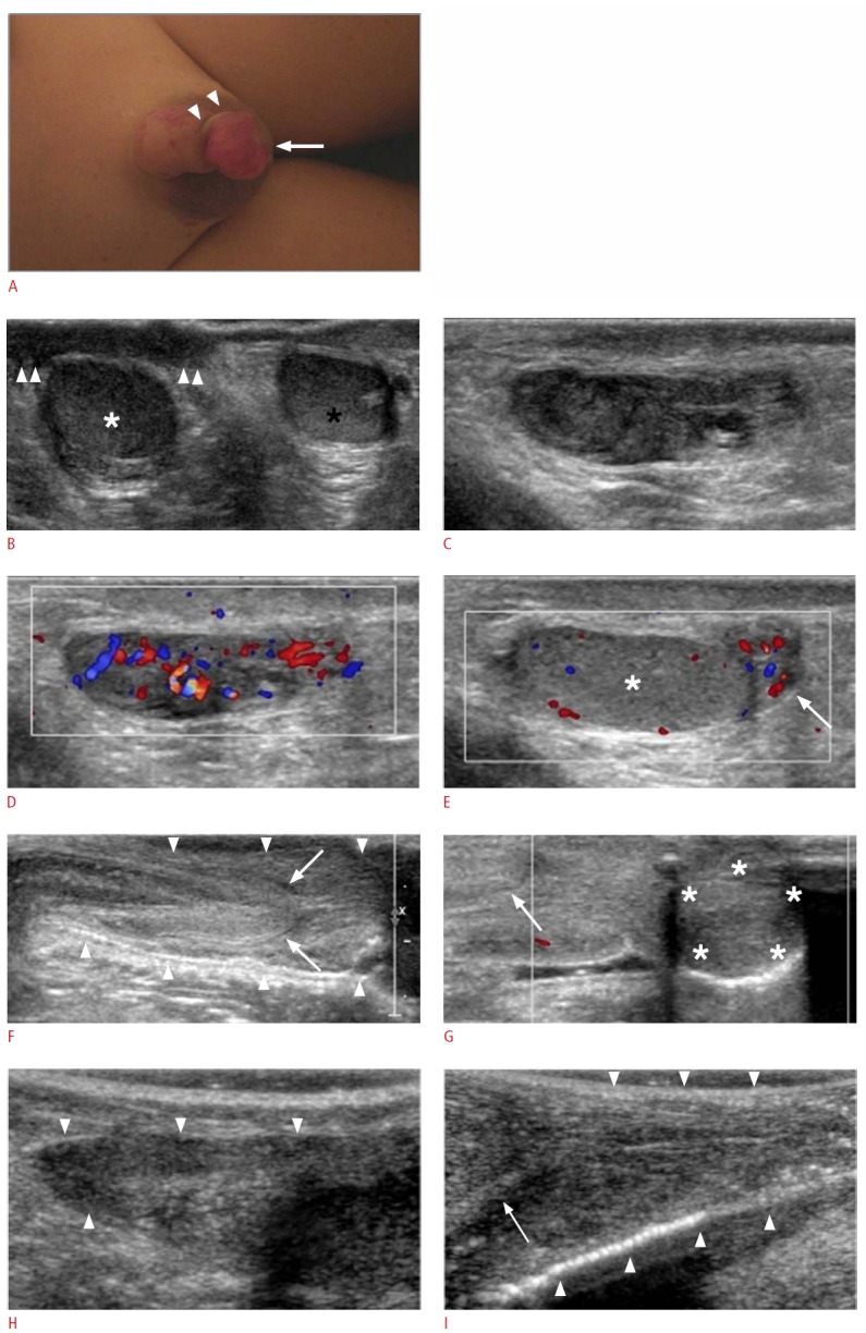
Fig. 1. A 5-year-old boy with symptoms of Henoch-Schonlein purpura.

A. Photograph shows the swelling of the right scrotum and the penis with a purpuric rash and a reddish mass-like lesion (arrow) at the penile tip. The reddish mass-like lesion causes skin twisting at the base (arrowheads) with mild deviation of the penile axis. B. Transverse ultrasonogram shows bilateral scrotal soft tissue thickening; more severe on the right side (arrowheads). White and black asterisks indicate the right and left testicles, respectively. C-E. Longitudinal grayscale (C) and color Doppler (D, E) ultrasonograms of the right epididymis show swelling with increased vascularity at the head and body (C, D). The tail of the right epididymis (arrow) also shows swelling with increased vascularity (E). The white asterisk indicates the right testicle. F, G. Longitudinal grayscale (F) and color Doppler (G) ultrasonograms of the penis show the swelling of the penile shaft and foreskin (arrowheads), and a well-defined hypoechoic mass-like lesion measuring about 1.4 cm×1.3 cm (asterisks) without vascularity on the penile tip. Arrows indicate the glans penis. H. Follow-up longitudinal ultrasonogram of the right epididymis (arrowheads) shows no swelling. I. Follow-up longitudinal ultrasonogram of the penis shows no swelling of the penile shaft and foreskin (arrowheads). The hypoechoic masslike lesion at the penile tip has disappeared. The arrow indicates the glans penis.
