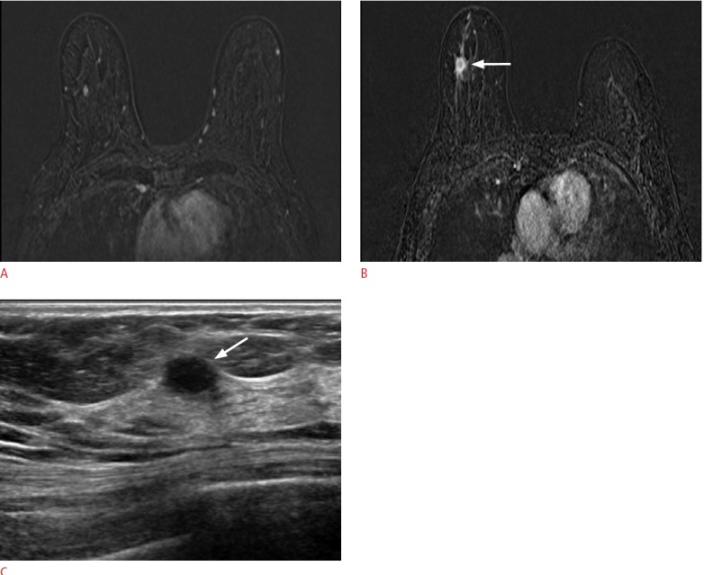
Fig. 3. A 57-year-old woman with cancer in her left breast.

A. A dynamic contrast-enhanced and subtracted T1-weighted axial image shows a small enhancing mass in her right breast. There were no correlated lesions seen on second-look ultrasonography (US). B. On follow-up magnetic resonance imaging performed 30 months later, a 1.5-cm irregular, enhancing mass (arrow) is detected at the same location in her right breast. C. US shows an oval hypoechoic mass (arrow) with an indistinct margin and confirmed it as invasive ductal carcinoma.
