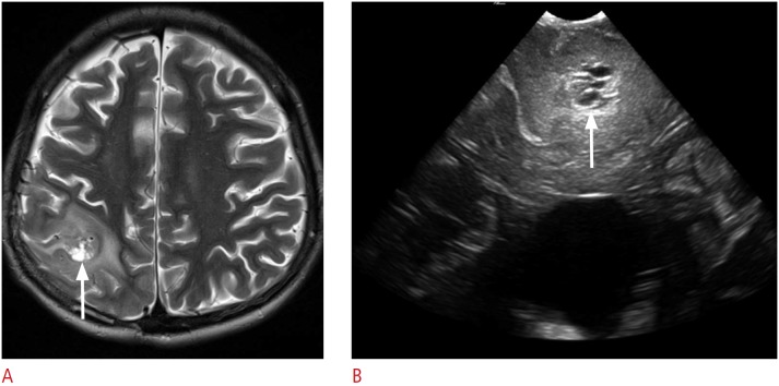
Fig. 5. A 13-year-old boy with a pathologically proven primitive neuroectodermal tumor.
A. Postoperative axial T2-weighted image shows an ill-defined hyperintense mass in the right parietal lobe with the surrounding edema. Note the central bright signal intensity area, which suggests intratumoral necrosis (arrow). B. Intraoperative ultrasonography demonstrates an ill-defined hyperechoic mass with a central low echoic portion (arrow) that corresponds to the central hyperintense area on the T2-weighted image. The mass and the surrounding edema are poorly differentiated. The pathologic report revealed a recurrent primitive neuroectodermal tumor.

