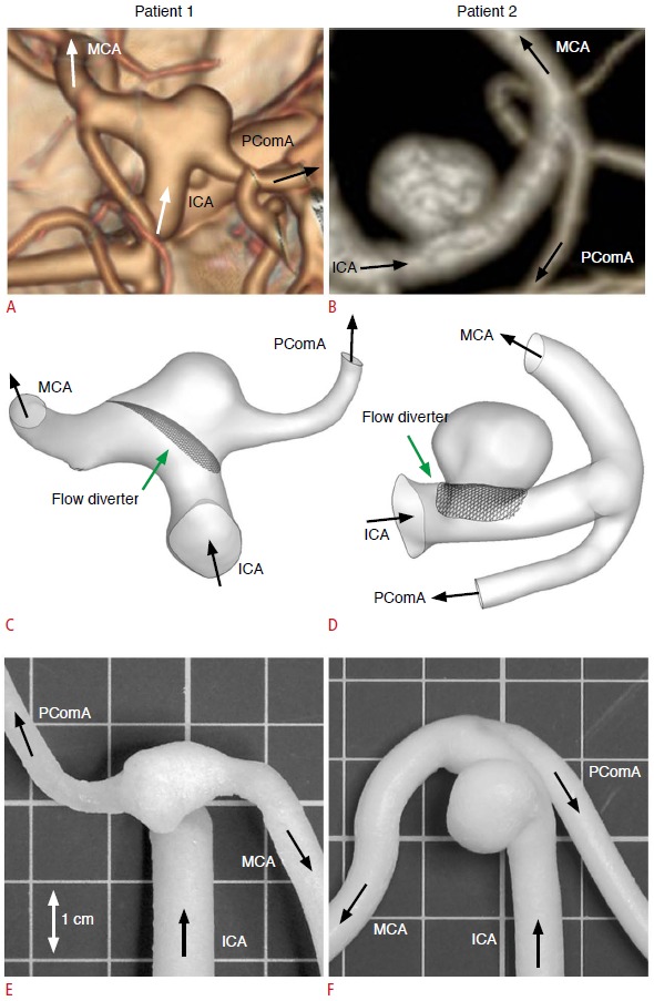
Fig. 1. Two intracranial aneurysms and the corresponding phantom models.

A, B. Computed tomography angiograms illustrate the configurations of a posterior communicating artery aneurysm in a 60-year-old woman (patient 1) (A) and a distal internal carotid artery aneurysm in a 71-year-old woman (patient 2) (B). C-F. These angiograms were used to generate computational fluid dynamics models (C, D) and physical phantom models (E, F). ICA, internal carotid artery; MCA, middle cerebral artery; PComA, posterior communicating artery.
