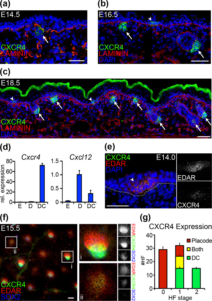
Figure 1. Cxcr4 expression in embryonic skin.
(a–c) Immunofluorescence staining for CXCR4 shows differential expression in epithelial placodes (arrowheads) and mesenchymal dermal condensates (arrows) of forming HFs at E14.5, E16.5, and E18.5. LAMININ labels basement membranes; Dapi highlights nuclei. (d) qRT-PCR of FACS sorted cells at E14.5 shows high Cxcr4 expression in dermal condensates (“DC”), compared to embryonic dermis (“D”) and epidermis (“E”). Cxcr4 ligand chemokine Cxcl12 is expressed in mesenchymal cells. (e) Immunofluorescence of E14.0 skin confirms CXCR4 expression in EDAR+ placodes of earliest forming HFs. (f) EDAR (placode marker) and SOX2 (condensate marker) expression highlight the stage- and compartment-specific localization of CXCR4 expression. In more advanced stage 2 follicles (i) CXCR4 co-localizes with SOX2. In early stage 0 follicles (ii) CXCR4 co-localizes with EDAR. (g) Quantification of CXCR4 localization in early forming HFs by stage at E15.0 (mean and SD of 3 embryos). CXCR4 expression switches from epithelial to mesenchymal cells. Dotted line denotes basement membrane. Scale bar = 50µm.

