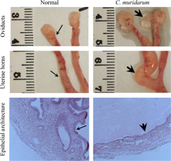
Fig. 4. Gross and microscopic pathology in C. muridarum challenged mice.

The histopathology was evaluated in mice on day 80 after C. muridarum or mock (PBS) challenge. The representative gross images from oviducts and uterine horns, and microscopic images (total magnification 25X) of uterine horn epithelium are shown. The normal (thin arrows) and dilated (thick arrows) oviducts and uterine horns are indicated. Additionally, the uterine epithelial architecture in H&E stained tissue sections displays abundant folds of the uterine epithelium in mock-challenged mice (thin arrow) versus a flattened, but continuous, epithelial lining (thick arrow) in C. muridarum challenged animals. Luminal exudate, with some inflammatory cells, is apparent in the tissues from infected mice.
