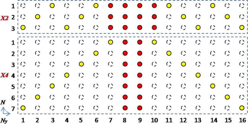
FIG. 1.
K-space sampling for PCLR. For both 2-fold (X2) and 4-fold (X4) undersampling, the half of the data (red dots) is acquired at the central k-space, while the other half (yellow dots) is acquired at the peripheral k-space. The peripheral sampling is circulant with respect to the DW dimension, and is repeated every 3 or 7 DW directions for X2 and X4 respectively. The k-space with the central data (red dots) and zero-filling elsewhere is used to generate the low-resolution phase estimate. Then with PCLR, the image reconstruction utilizes all the data (red and yellow dots) to reconstruct the DW images.

