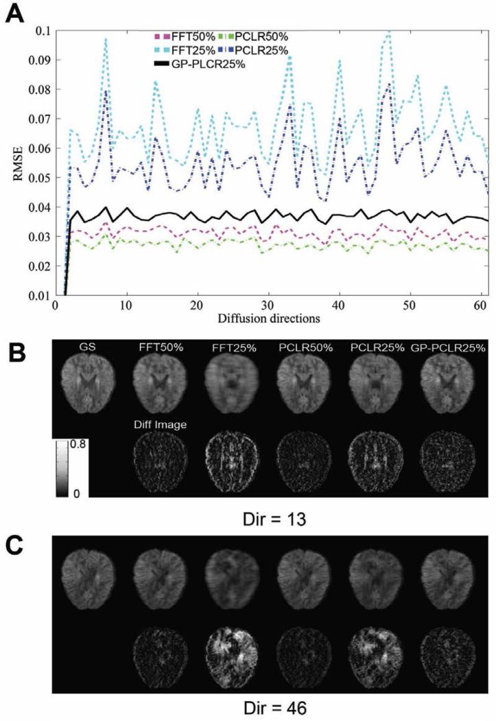
FIG.3.
The root-mean-squared-error (RMSE) of the reconstructed raw diffusion and the difference images across diffusion directions. The RMSEs between the gold standard and the reconstructed diffusion raw images of the whole brain across DW gradients were calculated and plotted in (A). Different methods (FFT, PCLR, GP-PCLR) and sampling rates (50%, 25%) were compared. It can be seen that GP-PCLR 25% showed less RMSEs than the PCLR25% and FFT25%. The representative axial slices of the raw diffusion images (upper row) and the difference images (lower row) at two diffusion directions (Dir=13, 46) were also shown in Fig.B, C. Except that GP-PCLR generates low RMSEs, the performances of the method across the directions are more consistent across directions than the corresponding PCLR25% and FFT25%, which is critical for the accurate estimation of DTI-derived measures. The difference images were magnified four times for easy visualization.

