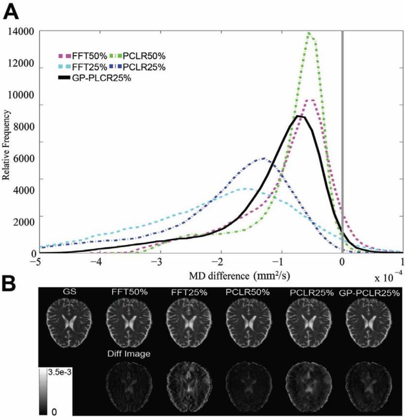
FIG.5.
The MD differences between the reconstructed images and the ground truth under various methods and undersampling rates. All reconstructed MD images tend to have overestimated MD in the most voxels in the brain. Compared to FFT25% and PCLR25%, GP-PCLR25% showed superior performance. The MD differences in a representative slice are shown in (B). The MD differences were magnified four for easy visualization.

