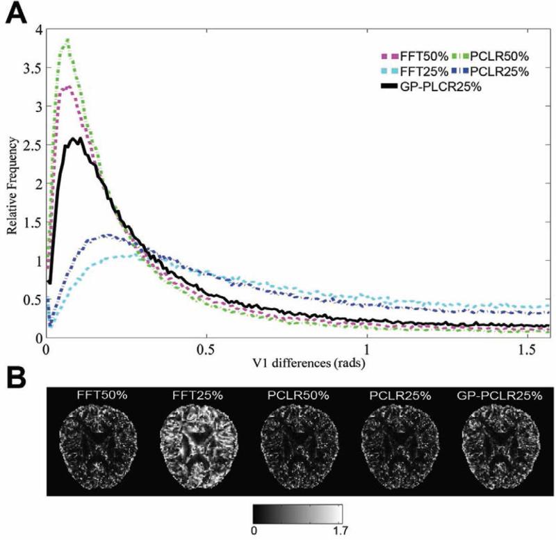
FIG.6.
The angular differences of the principal diffusion direction (V1) in the diffusion tensor model between the reconstructed images and the ground truth under various methods and undersampling. A narrow peak with high relative frequency centered near zero indicates more accurate reconstruction. GPPCLR significantly outperformed the other two methods (PCLR and FFT) under the same sampling rate (25%) (A). A representative slice of the angular differences is shown in (B). Generally, the estimation of the V1 is more accurate in the white matter than in CSF and gray matter. The unit of B is in radians

