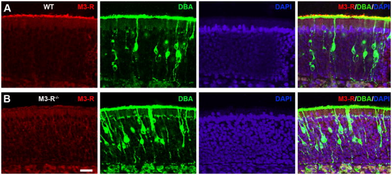
Figure 1. M3-R antibody stains the cilia layer of the olfactory epithelium in WT but not in M3-R knockout mice.

Coronal sections of the nose were stained with the M3-R antibody (red), DBA (green), and DAPI (blue) in WT (A) and M3-R knockout mice (B). Confocal images were taken at a single plane with z step = 1 μm.
