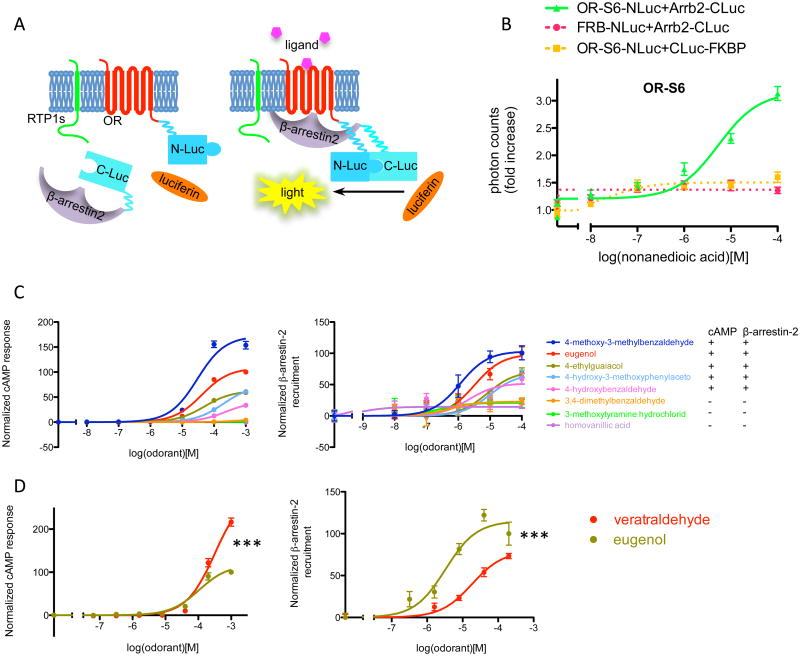
Figure 6. β-arrestin-2 recruitment assay for ORs.
(A) An overview. The luciferase gene is split into two fragments (N-Luc and C-Luc) and fused to the termini of OR and β-arrestin-2. RTP1S enhances the surface expression of many ORs (left). When ligand is present, the OR is activated and recruits β-arrestin-2. The association between the OR and β-arrestin-2 brings the two fragments of luciferase close to each other to form a functional enzyme, resulting in above-background light emission in the presence of luciferin. (B) Dose-response curve of β-arrestin-2 recruitment by a mouse OR, S6, stimulated by its ligand nonanedioic acid. Photon counts were normalized to background emission when no ligand is present to show the fold increase. Dose-dependent light emission was observed when OR-NLuc and Arrb2-CLuc were expressed, but not when either of them was substituted with control protein tagged with corresponding luciferase fragment (FRB-NLuc and CLuc-FKBP). (C) Dose-response curve of cAMP response and β-arrestin-2 recruitment for OR-EG when challenged with various chemicals, and summary of positive and negative responses. (D) Biased signaling towards cAMP pathway of the ligand veratraldehyde, as compared to eugenol. n=3, error bars indicate standard errors.

