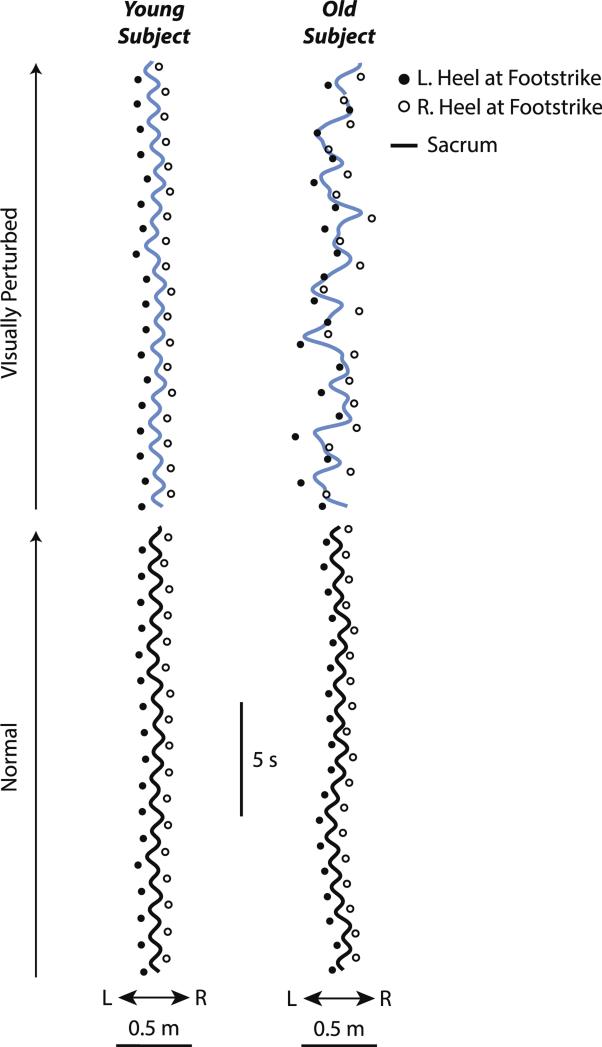
Fig. 2.
Relation between ML sacrum motion and lateral step placement during normal (black line) and visually perturbed (blue line) waking for representative old and young subjects. (For interpretation of the references to color in this figure legend, the reader is referred to the web version of this article.)

