
Figure 6. Visualization of exogenously added PFFs and p-α-syn aggregates formed from endogenous α-syn.
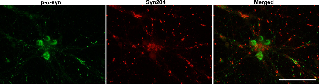
Neurons were fixed 14 days after treatment with PFFs and double immunofluorescence was performed using a rabbit antibody to p-α-syn generated in the Lee lab (green)14, and a mouse antibody, Syn204, that specifically recognizes the exogenously added human PFFs (red). There was minimal colocalization between the exogenous PFFs and the p-α-syn inclusions derived from endogenous α-syn. Scale bar = 50 µm.