
Figure 3.
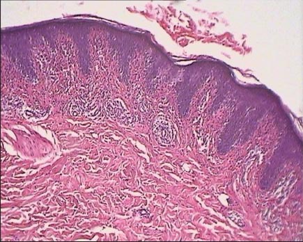
Histopathological feature of the patch stage of mycosis fungoides. Sections from the left arm (hypopigmented) patch show epidermotropism of atypical lymphocytes, mostly in basal layer and dermis, mild interstitial infiltrate of small lymphocytes seen (H and E×100)