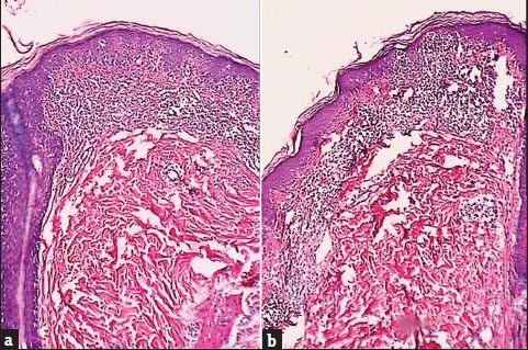
Figure 4.

Histopathological feature of mycosis fungoides. (a: Patch, b: Plaque.) Sections show epidermotropism of atypical lymphocytes in epidermis with focal Pautrieræs microabscesses. Upper dermis reveals prominent fibrosis with thick and wiry collagen bundles in haphazard directions intermingled with a moderately dense and band-like infiltration of atypical lymphoid cells (H and E×100)
