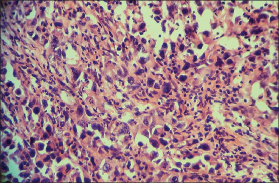
Figure 5.

Microphotograph of hematoxylin and eosin-stained section showing tumor cells with epithelioid morphology having plenty, pink cytoplasm, hyperchromatic nuclei, and prominent nucleoli with increased mitotic activity. Cells lacked melanin pigment. Original magnification: ×400
