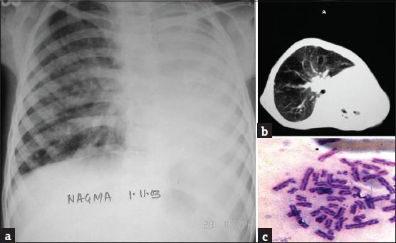
Figure 2.

(a) Chest radiograph showing left opaque hemithorax with mediastinal shift to the left suggestive of collapse of the left lung, (b) CECT (Contrast-enhanced computed tomography) of the chest showing complete volume loss of left lung with areas of bronchiectasis. The left main bronchus cannot be visualized, and, (c) High frequency of seen on chromosomal analysis of patient's blood sample
