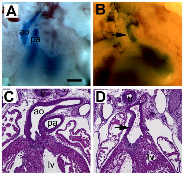
Fig. 1.
OFT defects in diabetic embryopathy. (A,B) Whole mount preparation of hearts at E15.5. The great arteries are labeled with blue dye. (C,D) Frontal sections of hearts at E15.5. (A,C) Non-diabetic controls. (B,D) Diabetic group. Arrows indicated single outflow trunk. ao, aorta; lv, left ventricle; pa, pulmonary artery; tr, trachea. Scale bar = 100 μm in A,B; 50 μm in C,D.

