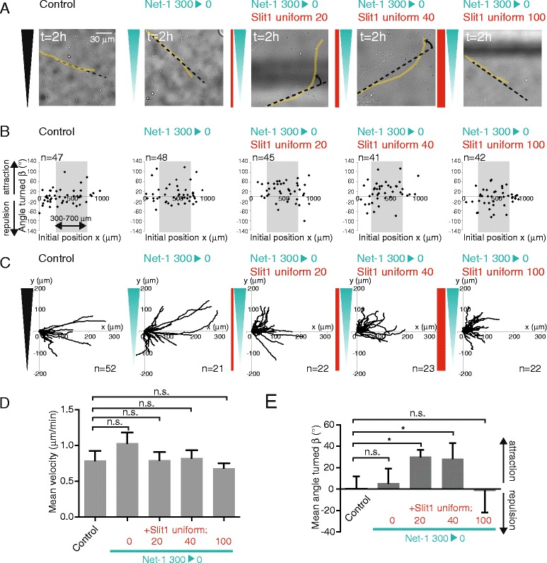
Figure 3.

Slit1 enables Netrin-1 attraction at low concentration. (A) Last brightfield images of typical growing axons. The growing neurite (yellow line), the initial orientation (dark dotted line), and the angle turned (rotating arrow) are shown. Bar, 30 μm. (B) Scatter plot of the angle turned versus the initial position (x). (C) Trajectory plots of growth cones in the different conditions, for initial positions between 300 and 700 μm. (D) The mean velocity (±SEM). n.s.: P > 0.05, Mann–Whitney test in which each condition is compared to the control. (E) The mean angle turned (β) (±SEM) for axons in the different conditions, for initial positions between 300 and 700 μm. Statistical differences are indicated *P < 0.05, Kruskal Wallis test with Dunn’s correction.
