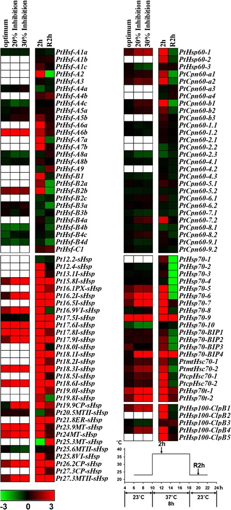
Figure 6.

Expression profiles of Populus Hsfs and Hsps under heat stress. Heatmap showing expression of Hsf and Hsp genes under heat stress. Microarray data (GSE26199) obtained from NCBI GEO database and our RNA-seq data (unpublished, see Methods) were used to be analyzed. In GSE26199, the expression changes of Hsf and Hsp genes under photosynthetic optimum (31.75°C, temperature producing the maximum net CO2 assimilation rate), 20% inhibition of optimum (38.4°C) and 30% inhibition of optimum (40.5°C) relative to baseline (22°C, the growth temperature) were analyzed in P. trichocarpa leaves. In our RNA-seq data, the expression changes of Hsf and Hsp genes during heat stress (2 h, 2 hours after heat treatment at 37°C; R2h, 2 hours after recovery from 37°C) were analyzed in the 5th internode of P. alba × P. glandulosa. The expression level of genes was determined based on the value of FPKM (Fragments Per Kilobase of exon per Million fragments mapped). Details of the FPKM are shown in Additional file 10: Table S9. Color scale represents log2 expression values, green represents low level and red indicates high level of transcript abundance. Blank represents the gene has no corresponding probe sets in the microarray data.
