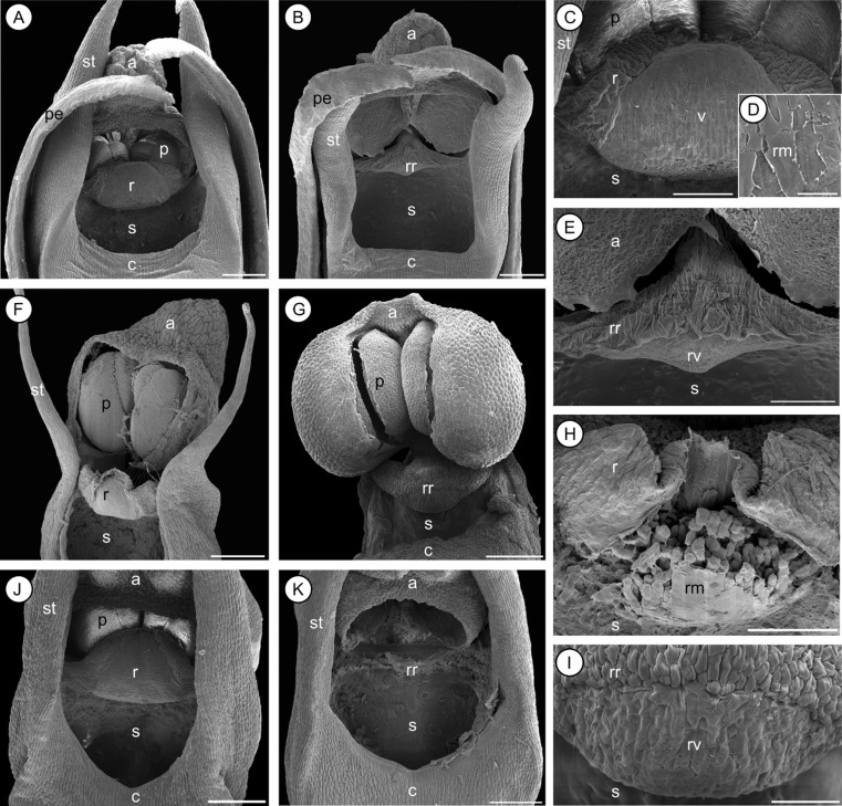
Figure 4.
Scanning electron microscopy micrographs of different gynostemium morphs (Types I versus II) observed in Bulbophyllum quadrifarium (A, B, C, D, E), Bulbophyllum erectum (F, G, H, I), and Bulbophyllum occultum (J, K). A, B, gynostemia of B. quadrifarium (Type I and II, respectively). C, rostellum of Type I; the central-lateral part has marked epidermal-cuticular foldings, whereas the anterior part is comprised of the half-oval viscidium. D, close-up of the viscidium surface; a smooth, extracellular rostellar membrane covers the viscidium cells. E, rudimentary rostellum and viscidium of Type II. F, G: gynostemia of B. erectum (I, II); note the lack of stelidia in G. H, rostellum of Type I; the disrupted rostellar membrane only partly covers the viscidium cells. I, rudimentary rostellum and viscidium of Type II. J, K: gynostemia of B. occultum (I, II). a, anther; c, column; p, pollinia; pe, petal; r, rostellum; rm, rostellar membrane; rr, rudimentary rostellum; rv, rudimentary viscidium; s, stigmatic cavity; st, stelidium; v, viscidium. Scale bars = 0.2 mm (A, B, F, G, H); 0.1 mm (C, E, H); 0.01 mm (D); 0.05 mm (I). All micrographs by A. Gamisch and U. Gartner (only F).

