
Figure 5.
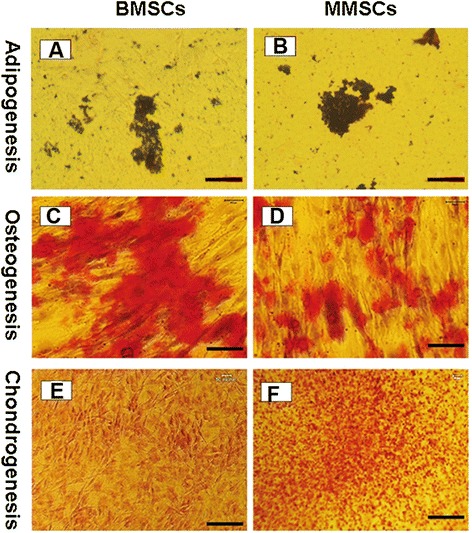
Histochemical staining of differentiated cells and semi-quantification of the extent of cell differentiation. Both BMSCs and MMSCs were able to differentiate into adipocytes (A, B), osteocytes (C, D), and chondrocytes (E, F), as shown by the accumulation of lipid droplets, proteoglycans and calcium deposits on cell surfaces. However, higher extent of osteogenic differentiation in BMSCs was evidenced by the most positive staining areas through Alizarin Red S assay. Conversely, much higher potential of chondrogenic differentiation in MMSCs was verified by Safranin O staining. Note that each experiment was repeated three times using five different donors. (P < 0.05) (Magnification of microscopy: 20×) (Bar: 50 μm).