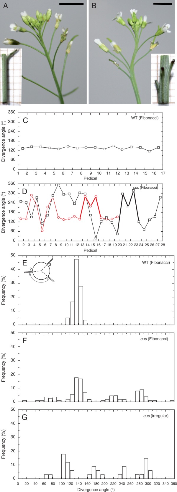
Fig. 1.

Pedicel phyllotaxis. Inflorescence shoots of WT (A) and cuc2 cuc3 (B) plants. Insets show magnified views of elongated shoot fragments with one (A) or several (B) pedicels departing from the stem (the background grid is 1 × 1 mm). Note the uneven distribution of pedicels along the stem, and fusions between the stem and pedicels in the mutant. Scale bars = 5 mm. (C, D) Graphs representing the divergence angle measured between consecutive pedicels of one exemplary WT (C) and two cuc2 cuc3 (D) shoots, indicated by different colours. Line segments representing exemplary M-motifs (D) are thickened. (E–G) Histograms of divergence angles measured between pedicels at levels where they separate from the stem for WT (E) and cuc2 cuc3 (F, G) plants.
