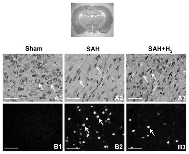
Figure 3.
Nissl staining was performed on brain sections obtained from the sham, subarachnoid hemorrhage (SAH), and hydrogen (H2) treatment groups. Neurons in the cerebral cortex at 24 hrs after SAH (n = 3) are shown in upper panels. Intact neurons could be seen in sham animals. Arrows indicate normal neurons (A1). Numerous foci of neuronal damage were present in the cerebral cortex in SAH animals. Arrows indicate deformed neurons (A2). Hydrogen treatment prevented the appearance of injured neurons and more positive stained cells and better cellular shape were observed (A3). The bottom panels show the results of terminal deoxynucleotide transferase-mediated dUTP nick-end labeling staining after 24 hrs after SAH (n = 3). No positive staining was detected in the sham group (B1). SAH rats presented massive terminal deoxynucleotide transferase-mediated dUTP nick-end labeling-positive cells in the cerebral cortex (B2), whereas evident reduction of terminal deoxynucleotide transferase-mediated dUTP nick-end labeling-positive cells could be seen in the SAH + hydrogen group (B3). Arrows indicate cells that are positive. Scale bars represent 100 μm.

