
Fig 8.
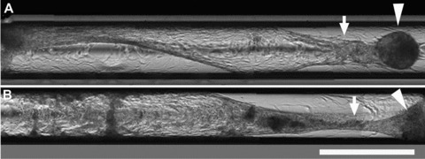
Tubulogenesis in capillaries. HPTCs are seeded into glass capillaries with an inner diameter of 580 μm. (A) and (B) show two different capillaries containing HPTCs imaged ∼2 weeks after seeding. Several images were stitched together in order to cover a larger area. Initially after seeding, monolayers covering the inner walls of the capillaries are formed. The monolayer is still intact in the left half of the lower capillary (B). Myofibroblast aggregates appear after monolayer formation. The monolayer is then rearranged and detached from the capillary walls, and tubules are formed within the capillaries (marked by arrows), which are attached to myofibroblast aggregates (marked by arrowheads). Scale bar: 1 mm.