
Fig 1.
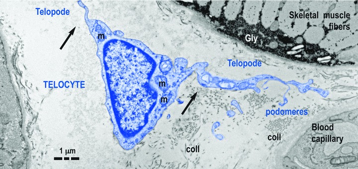
Telocyte (TC) in human skeletal muscle interstitium: two striated fibres and a blood capillary (transmission electron microscopy). Cellular body of TC (digitally coloured in blue) has a thin layer of cytoplasm with few mitochondria (m) around nucleus. Telopodes (Tps) have a narrow emergence (arrows) from cellular body. Tps have a sinuous trajectory, podomeres (extremely thin segments, below the resolving power of light microscopy) and podoms (dilated portions). coll: collagen fibres.