
Fig 3.
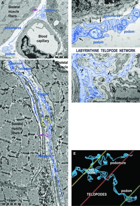
Ultrastructural features of Tps in rat (A) and human (B–D) striated muscle. (A) One Tp in between a blood capillary and two striated muscle fibres (m, mitochondria). (B) Details of a podom, which accommodates mitochondria (m), endoplasmic reticulum (ER) and caveolae (black arrows). (C, D) Tps with bifurcations and a labyrinthine configuration among striated muscle cells. The yellow circle (C) and black arrows (D) indicate close contacts between Tps of different TC. (E) Computer simulation of a 3D representation of a TC outside the tissular environment. Note that various sections (coloured in yellow, orange and green) contain different segments of Tps.