
Fig 6.
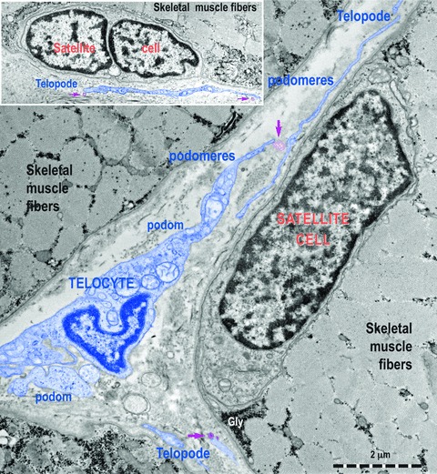
Transmission electron micrographs show TC with Tps, podoms and podomeres in between muscle fibres. Note the typical appearance of satellite cells. TC (digitally blue coloured) are positioned in the close vicinity of satellite cells. Two ultrastructural features are remarkable: the close spatial relationships of Tps with satellite cells and the fact that these Tps release shed vesicles (purple arrows). This may indicate that a transfer of chemical information flow from TC to satellite cells.