
Fig 9.
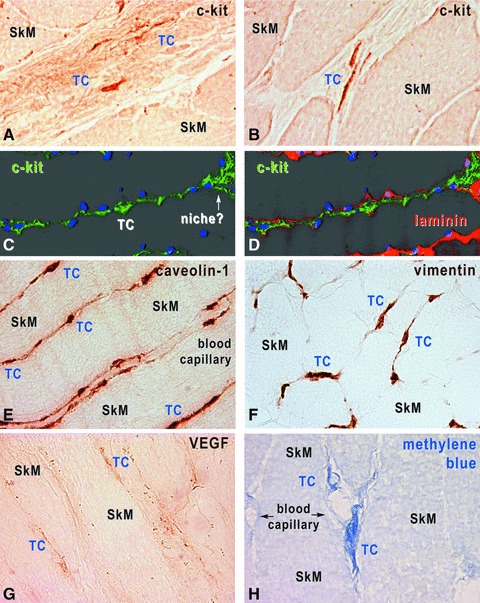
Human skeletal muscle, immunohistochemistry with HRP conjugated antibodies on cryosections (A, B, E, H) and immunofluorescence-confocal microscopy (C, D). TCs are located within interstitium, and express c-kit (A–D), caveolin-1 (E), vimentin (F) and VEGF (G). TCs were also revealed by methylene blue vital staining (H). In C and D, TCs are identified by c-kit expression (green), the basal lamina by laminin expression (red) and nuclei are stained with DAPI (blue). Original magnification 1000×.