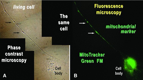
Fig 12.

Mouse skeletal muscle in cell culture. The same cell was analysed by (A) phase contrast microscopy and (B) fluorescence microscopy after labelling the mitochondria of living cells with MitoTracker Green FM. Mitochondria are concentrated around the cell nucleus and within the podoms (arrows). Photographic reconstruction; original magnification: 1000×.
