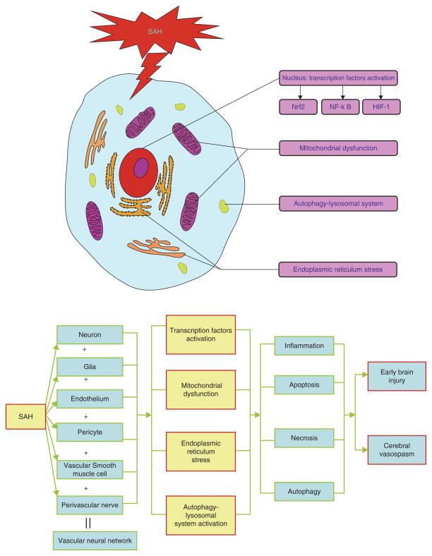
Fig. 1.
The functional disturbance of organelles in the pathogenesis of SAH. The components of the vascular neural network of the brain, including neurons, glia, endothelium, pericytes, vascular smooth muscle cells, and perivascular nerves, all suffer from SAH-induced injuries. The dysfunctions/functional alterations of the organelles that take place are in the transcription factors (e.g., Nrf2, NF-κB, and HIF-1), mitochondrial dysfunction, endoplasmic reticulum stress, and the autophagy–lysosomal system. These pathophysiologic cascades play a critical role in inflammation, apoptosis, necrosis, and autophagy in brain parenchyma after SAH

