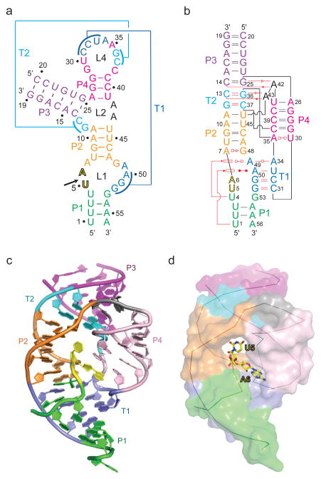
Figure 1. Schematics of the secondary structure and the 2.9 crystal structure of the dU5-containing env22 twister ribozyme.
a, A schematic of the secondary fold of the env22 twister ribozyme composed of a 19-mer substrate strand containing dU5 at the U5-A6 cleavage site. The sequence is color-coded according to helical segments observed in the tertiary structure. Two proposed pseudoknot segments are labeled T1 and T2. b, A schematic of the tertiary fold observed in the crystal structure of the env22 twister ribozyme. The red lines indicate tertiary pairing. The geometric nomenclature and classification of RNA is adopted from reference 33. c, A ribbon view of the 2.9 Å structure of the env22 twister ribozyme color-coded as shown in panels a and b. d, The same view as in panel c but with the RNA in a surface representation except for the U5-U6 step, which is shown in a stick representation.

