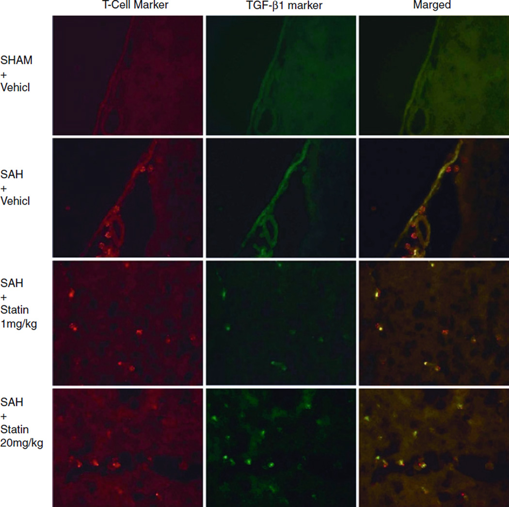
Fig. 3.
Representative photographs of double-fluorescence labeling for transforming growth factor (TGF) β1 and T cells in the brain slices mounted on histological glass slides. The mouse anti-T-cell maker indicated T cells are not visualized in the subarachnoid space following sham surgery but become present following subarachnoid hemorrhage (SAH). The TGF-β1 expression by leukocytes was detected following treatment with simvastatin with the use of rabbit anti-TGFβ-1 antibodies. Samples collected 24 h after SAH

