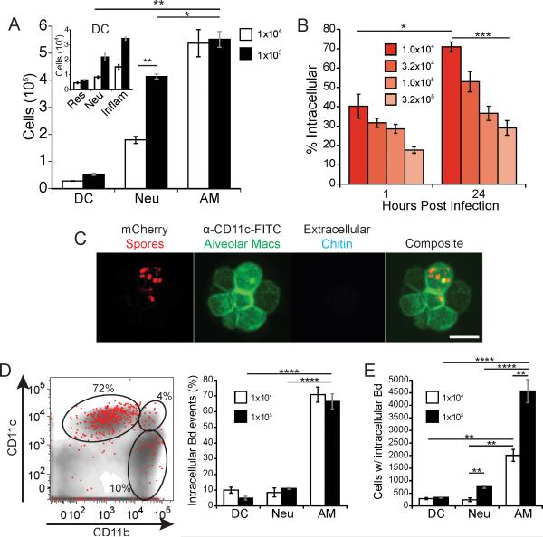
Figure 1. The majority of spores reside in alveolar macrophages early during infection.
Mice were infected with mCherry 14081 spores by intubation. (A) At 24 hours post infection, lung homogenates were analyzed for the total number and lineage of leukocytes. The gating markers and strategy are described in the Methods and illustrated in supplemental Fig. S1. Alveolar macrophages (AM); neutrophils (Neu), dendritic cells (DC). DC subsets (inlay) are resident (Res), neutrophil-derived (Neu), and inflammatory (inflam). (B) The proportion of total spores (mCherry+ events) in the lungs that are intracellular (Uvitex 2B−) at 1 and 24 hours post infection was measured at a range of inocula. Results are representative of three experiments each with five mice/group. (C) Intracellular residence of spores within alveolar macrophages from lung homogenates of mice infected with mCherry spores for 12 hours. Alveolar macrophages were identified with anti-CD11c-FITC. Uvitex 2B stain of chitin was used to determine if the spores were extracellular (only extracellular spores bind the dye). Bar in lower right panel = 25 microns. (D) Distribution of intracellular spores (mCherry+, Uvitex 2B−) 24 hours after mice were infected with 1×10⁵ spores. A flow plot of a representative mouse illustrating intracellular spores (red dots) overlaid on total leukocytes (gray) with representative gates for alveolar macrophages (top left), DCs (top right), and neutrophils (bottom right). Distribution of spores within leukocytes (left panel) is based on the gating strategy shown in the Fig. S1; group mean±SEM analyzed with FACS (right panel). (E) The total number of lung leukocytes that contain spores 24 hours post infection. Results shown in panels D and E are representative of 3 experiments with 5 mice per group. *P<0.05, **P<0.01, ***P<0.001, ****P<0.0001.

