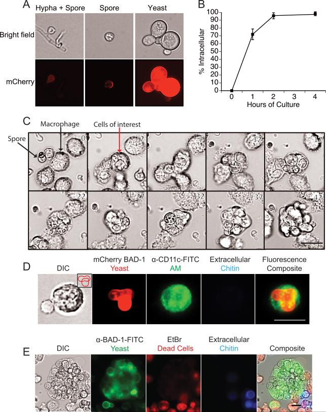
Figure 2. B. dermatitidis spores survive and replicate as yeast in alveolar macrophages.
(A) Bright field and fluorescence microscopy of spores and yeast of reporter strain 14081 that upregulates mCherry under the control of a yeast phase-specific BAD-1 promoter. mCherry fluorescence is quantified by FACS in Fig. 6A. (B) Uptake of mCherry spores by macrophages in vitro. Spores were cultured with primary alveolar macrophages at an MOI of 0.2. Uptake was quantified over 4 hours and analyzed by FACS. Extracellular spores stained Uvitex 2B+. Results are the mean of triplicate wells and representative of 3 experiments. (C) Live imaging of mCherry 14081 spores cultured with primary alveolar macrophages at a MOI of 0.2. Images shown are every 5 hours. The full movie is available online. (D) Mice were infected with mCherry 14081 spores and 3 days later BAL fluid was collected and stained with anti-CD11c-FITC to identify alveolar macrophages and Uvitex 2B to distinguish intracellular and extracellular yeast. Inlay in the left panel shows a budding yeast cell overlying another yeast. (E) Alveolar macrophage cell line AMJ2-C11 was cultured for 48 hours with 14081 yeast on coverslips. Cultures were treated with Uvitex 2B to stain the extracellular yeast, and with ethidium bromide to ascertain macrophage membrane integrity and cell viability. Cultures were fixed and then permeabilized to identify yeast with anti-BAD-1-FITC. Bar in lower right panels = 25 microns

