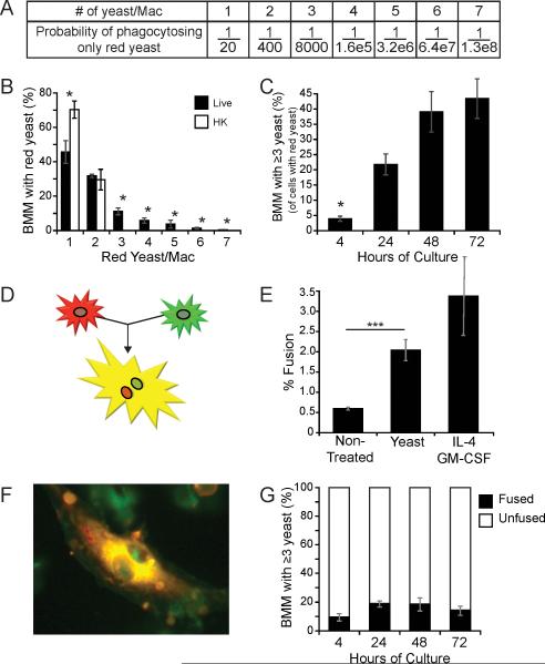
Figure 3. Intracellular replication by B. dermatitidis yeast.
(A) Probability of only red yeast being phagocytosed when they comprise 1/20th of the yeast inoculum. (B) BMM were cultured for 24 hours with two yeast strains, one expressing red fluorescent protein and the other, green fluorescent protein (ratio of 1:20, respectively); both strains in an assay were either live or heat-killed. The number of yeast per macrophage was enumerated for the BMM that contained only red yeast. 500 yeast-containing macrophages were enumerated per condition; results are the mean±SEM of five experiments. (C) BMM containing ≥3 red yeast were enumerated. 500 yeast-containing macrophages were counted per time point; results are the mean±SEM of three experiments (D) The fusion of a red macrophage with a green one results in a yellow, multinucleated giant cell. (E) Fusion between PKH26 (red) and CMFDA (green) stained BMM (1:1) was enumerated for cells cultured 24 hours with or without yeast (strain 26199) or with IL-4 and GM-CSF (control). Results are the mean±SEM of 4 experiments in which >400 macrophages were counted per condition. (F) A fused, yellow, multinucleated giant cell from a culture with yeast that does not contain intracellular yeast. (G) Red and green BMM were cultured with yeast over time. The proportion of fused BMM with ≥3 intracellular yeast is depicted. Results are the mean±SEM of 3 experiments in which >400 macrophages were counted per condition. *P<0.05, ***P <0.001.

