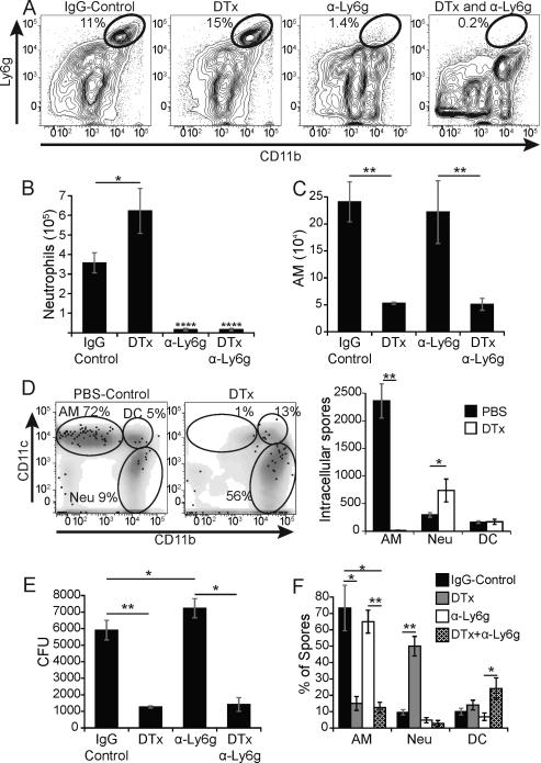
Figure 5. Neutrophils do not account for reduced lung CFU in CD11c depleted mice.
CD11c-DTR mice were treated with rat IgG, 100 ng DTx i.p., anti-Ly6g antibody i.v. or both DTx and antibody. Mice were infected with mCherry 14081 spores and lung homogenates were analyzed 2 days later. (A) Flow plots showing the proportion of lung neutrophils in representative mice. (B) Neutrophil numbers were quantified by FACS and hemocytometer count. (C) The number of alveolar macrophages (AM) in mice given DTx alone or together with neutrophil depleting antibody. (D) Flow plot of the distribution of intracellular spores (mCherry+, Uvitex 2B−) denoted as black dots with respect to leukocytes (gray) and alveolar macrophages (AM; top left gate), DCs (DC; top right), and neutrophils (Neu; bottom right) in representative PBS- vs. toxin-treated CD11c-DTR mice (left panel). The right panel shows the number of intracellular spores associated with each cell type. (E) Lung CFU in mice corresponding to panels A-C. (F) The distribution of spores among leukocytes in depleted or control mice evaluated by FACS. All results are representative of 3 independent experiments with 5 mice/group. *P<0.05, **P<0.01, ****P<0.0001.

